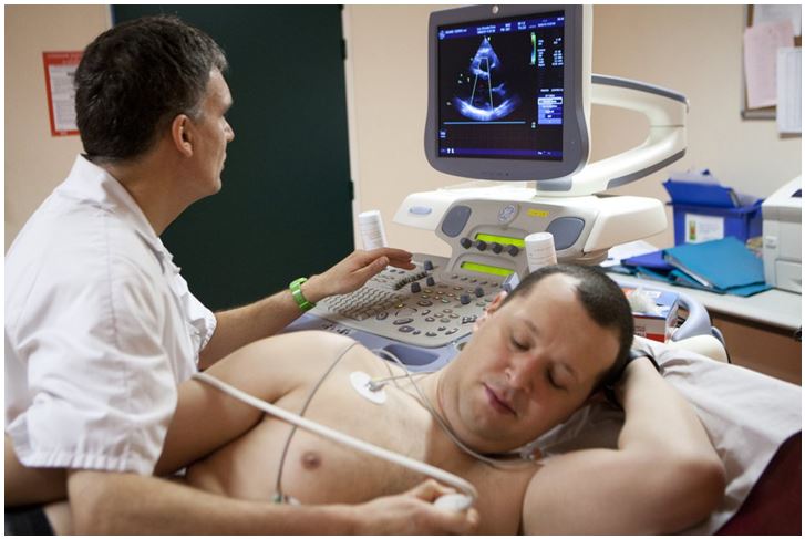
2D echocardiography (also known as a transthoracic echocardiogram) is a diagnostic test used to evaluate the structure and function of the heart. It uses high-frequency sound waves (ultrasound) to create detailed images of the heart and its chambers, valves, and blood vessels.
Dr.
Ravi Bhushan, a physician specializing in the diagnosis and treatment of heart
conditions, may use 2D echocardiography to evaluate a patient's heart function
and identify any abnormalities or problems.
During the test, a gel is applied to the patient's chest and a small transducer (handheld device) is moved over the chest to produce the images of the heart. The patient will lie on an examination table and the transducer will be moved over the chest to capture images of the heart from different angles. The test is non-invasive, painless, and does not use ionizing radiation.
2D echocardiography can help Dr. Bhushan to evaluate the heart's chambers, valves, and blood vessels, detect any structural abnormalities, and assess the heart's function and blood flow. The test can also help identify any problems with the heart's valves, such as stenosis (narrowing) or regurgitation (leakage).
It is also used to evaluate conditions such as heart failure, hypertension, and to assess the effectiveness of any treatment that is being used for these conditions.
It's important to note that 2D echocardiography is a safe
and painless diagnostic test that can provide valuable information about the
structure and function of the heart. However, it's always recommended to
consult with the physician in charge to understand the reason of the test and what
should be expected.
Copyright @ Dr. Ravi Bhushan Designed By Persistent Infotech